But one of the major advantages was the ability of Nanolive Imaging to capture several biological phenomena and objects at the same time, enabling researchers to observe organelle rotation and other complex cellular dynamics involving multiple subcellular structures.


The label-free, non-invasive imaging technique meant the team could observe the cells over an extended period of time. Taken together, the data paints a detailed picture of a cell’s structure and chemical composition, allowing scientists to precisely calculate dry mass, morphology, cell membrane dynamics and other features. They then quantified the observed phenomena using software specially designed to sift through the mass of raw imaging data.

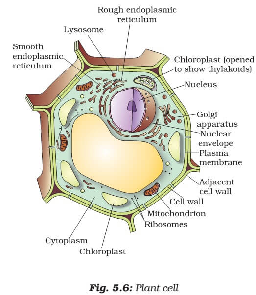
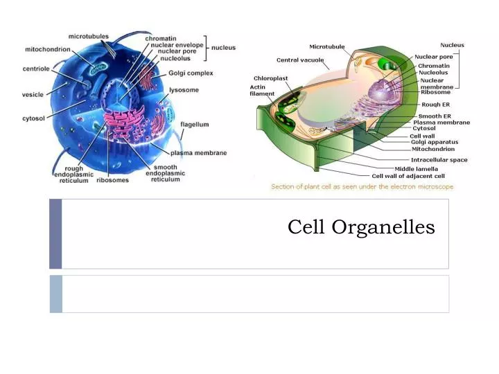
In this paper published in PLOS Biology in December 2019 by using Nanolive’s 3D Cell Explorer-fluo, researchers from EPFL could observe changes in cell size during division, organelle movements, and the formation of tiny lipid droplets – all over an extended period and without damaging the cell. Organelles of an animal cell showing different components present in a eukaryotic cell.


 0 kommentar(er)
